"Human Kidney Description, Location, Functional Parts,
Etc."
Kidneys are located at the level of the 12th
thoracic and first two lumbar vertebrae. The right kidney is a little lower
than the left because the liver presses it down. The superior pole of the
right kidney is crossed from behind by the 12th rib. In the case of
the left kidney, the 12th rib divides it into two parts: a
superior, smaller part (1/3), and an inferior, larger part (2/3). The kidneys
are related behind to the quadratus lumborum muscle, the psoas muscle, and the
lumbar part of the diaphragm. They have anterior and posterior surfaces. The
anterior surface is a little lateral; the posterior surface is a little
medial. So, they're not exactly in the frontal plane. They have superior and
inferior poles. Above the superior pole, sit the suprarenal glands. On the
medial border (it is concave), we have the hilus. The lateral border is
convex.
The kidneys are fixed to the abdominal cavity by three
capsules. The most important is the outermost capsule which is called fascia
renalis. This fascia layers the anterior surface of the kidneys, continues
to the posterior layer at the lateral margin of the kidneys, and continues to
the posterior layer above the kidneys. So, it is a closed capsule superiorly and
laterally, but it is open inferiorly and medially. Medially, the anterior layer
passes in front from the aorta and inferior vena cava, continues to the other
side (anterior surface) and laterally sides of the kidneys continues to the
posterior layer behind the aorta and inferior vena cava.
The anterior layer of the fascia renalis is fused with the
parietal peritoneum. The posterior layer is fused with the transverse fascia
(fascia transversalis) which is the innermost layer of the abdominal wall.
Between the two layers, the middle capsule, the adipose capsule, fills
the space between the two layers of the fascia.
The innermost capsule is the fibrous capsule which is
directly on the surface of the kidney. Between the fibrous capsule and the
renal fascia, there are connective tissue fibers through the adipose tissue. So,
finally, the renal fascia is connected to the fibrous capsule and the fibrous
capsule to the kidneys. The renal fascia is connected to the abdominal wall by
the transverse fascia and parietal peritoneum. This is the most important
support for the kidneys.
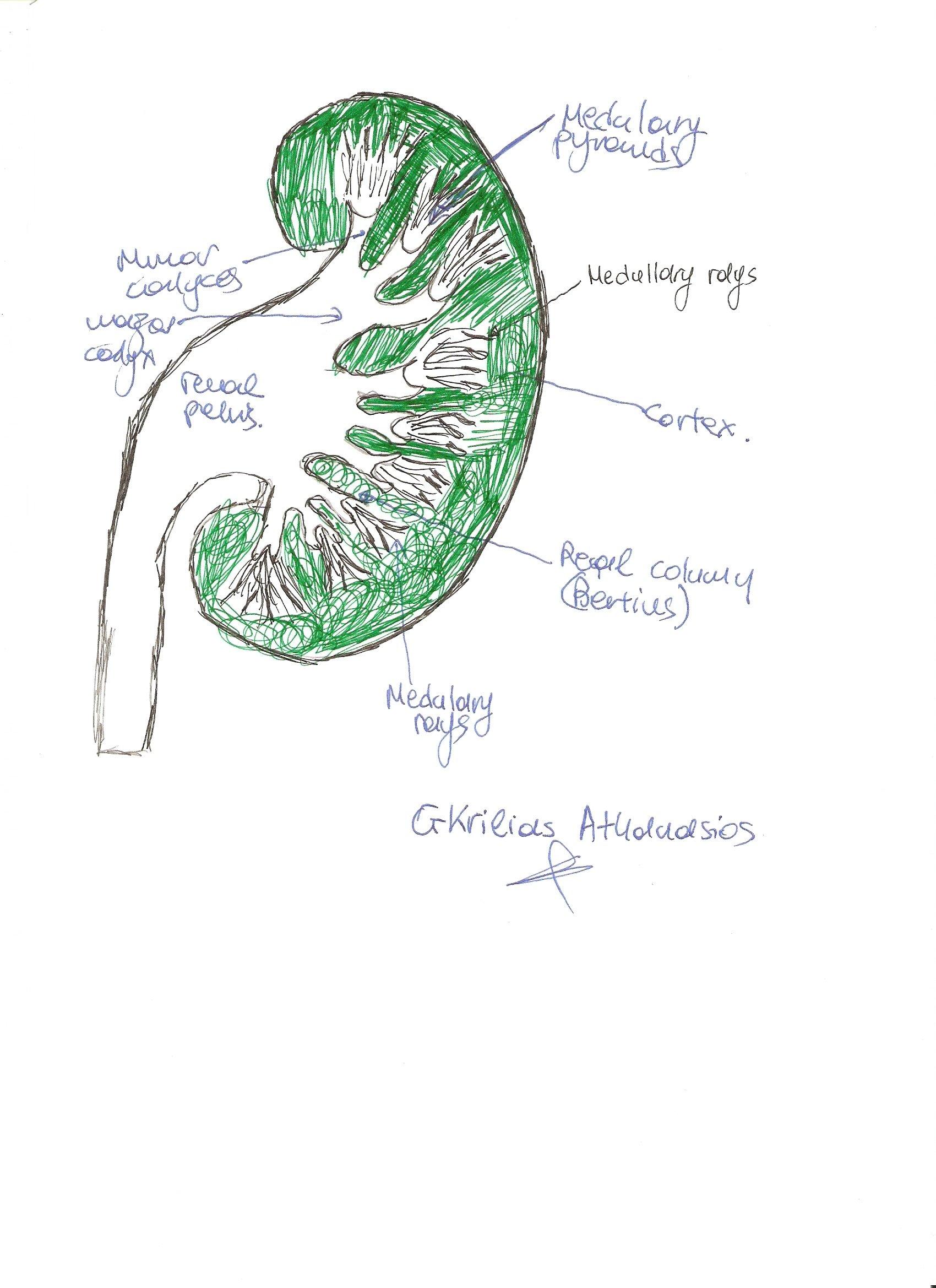
On the medial margin of the kidney, the hilus opens
into the sinus of the kidney. The sinus is a cavity of the kidney which is
surrounded by the parenchyma of the kidney (parenchyma: functional tissue of an
organ). The sinus contains the lesser calyces, the greater calyces, the branches
of the renal artery and vein (with loose connective tissue and fat), and the
pelvis which continues into the ureter. The adipose capsule continues into the
sinus.
Sinus = cavity.
Hilus = entrance of this cavity.
Pelvis = one of the structures of the cavity that belongs to
the urine system, collecting the calices.
The ureter starts at the level of the hilus and is the
inferior posterior structure of the hilus. The anterior-posterior order of
structures is vein-artery-ureter.
If you make a frontal section through the largest plane,
you will see that the outermost layer is the fibrous capsule on the surface.
The next layer is called the cortex cortices (right below the fibrous
capsule). Beneath this, the cortex forms the cortical columns between the
medullary pyramids. Inside the cortex, there are striations called
medullary rays (stria medullaris corticis). The cortex continues into the
medulla as cortical columns (columnae renalis or Bertin's columns).
The next part of the kidney is the medulla, forming the medullary
pyramids. The apeical (papillary) openings are situated on the minor calyx. On
the surface of the apex, there are tiny openings for the papillary ducts. It
is called lamina cribrosa because of these openings.
 The kidney
develops from lobes. One original lobe was one pyramid and a half of the
cortical column (renal column). Approximately 25-30 original lobes have fused
with each other and open to one minor calyx. Minor calyces are about 8-10 in
number. Three minor calyces open to one major calyx, so there are about three
major calyces that open into the renal pelvis.
The kidney
develops from lobes. One original lobe was one pyramid and a half of the
cortical column (renal column). Approximately 25-30 original lobes have fused
with each other and open to one minor calyx. Minor calyces are about 8-10 in
number. Three minor calyces open to one major calyx, so there are about three
major calyces that open into the renal pelvis.
Pelvis renalis: it is the dilated first part of the ureter
which is collected from the three major calyces and continues into the ureter.
It is located in the sinus of the kidney. The other name of the pelvis is pyelos,
and the infection inside is called pyelonephritis.
Renal arteries come from the abdominal aorta, belonging to
the paired visceral branches of the abdominal aorta. It enters the kidney
through the hilus and divides into interlobar branches which run in the middle
of the Bertin's columns (renal). The renal artery first divides into two
main groups of arteries, one in front of the main plane and one behind. From
these main arteries, we have the additional interlobar branches. If you cut the
kidney through the largest plane, you will not cut the main arteries, because
one is in front of the plane and the other behind.
The left and right renal veins drain to the inferior vena
cava. The left renal vein passes in front of the abdominal aorta across the
midline because the inferior vena cava is on the right. As a consequence of this
asymmetry, the left renal vein receives the
left testicular vein or
ovarian vein, but the right does not. Usually, an additional renal artery
(accessory) supplies the superior or inferior pole of the kidneys.
Muscles Related to the Kidney:
Quadratus lumborum, psoas major (hilus), and lumbar part of
the diaphragm.
The muscle that fills the iliac fossa is the iliacus that
inserts to the lesser trochanter of the femur. Its function is flexion of the
hip joint (it is the main flexor).